For an mRNA therapeutic, a plasma concentration-time curve can be perfectly accurate and still leave the most important pharmacological question unanswered: how much functional protein was produced in the cells that needed it? Circulating mRNA, tissue-associated mRNA, translation-competent intracellular mRNA and the encoded protein can all describe different parts of the same dose-to-response chain.
In my previous article, I looked at how those uncertainties affect first-in-human dose selection. Here, the focus is one step earlier: what should we treat as exposure, which measurements genuinely inform that exposure, and how much mechanism does a useful PK/PD model need?
What does “exposure” mean for an mRNA therapeutic?
For a lipid nanoparticle (LNP)-delivered mRNA therapeutic, the measurable quantities may include individual lipid components, circulating mRNA, tissue-associated mRNA, intracellular mRNA and the encoded protein. Each describes a different part of the path from administration to effect. None should be substituted for another without evidence that the relationship is sufficiently dependable for the intended use.
The clinical mRNA-1944 study illustrates the distinction unusually clearly. This product encoded the heavy and light chains of a chikungunya-neutralising antibody. Reported serum terminal half-lives were approximately 7.3 hours for the ionisable lipid in the single-dose analysis, 83.5 and 86.2 hours for the two mRNAs, and 69 days for the antibody. These are analyte-specific measurements in circulation. The mRNA results do not measure its lifetime in the cytoplasm, and the lipid result does not establish the persistence of an intact nanoparticle. PMID: 34887572
The same protein concentration can also occupy two positions in the pharmacological chain. Relative to administered mRNA, protein production is a downstream response; relative to the protein’s target, its concentration is an exposure variable. Being explicit about that relationship is more useful than insisting on a single boundary between PK and PD.
From distribution to productive delivery
I find it useful to separate three questions. Where does the administered material go, how much translation-competent mRNA becomes available in the relevant cells, and how much functional protein is subsequently present at its site of action?
The first concerns distribution: concentrations or amounts of measured components in blood and tissues. The second concerns what I will call productive intracellular exposure, meaning the amount and persistence of translation-competent mRNA in the appropriate cytoplasmic compartment. This is a descriptive term, and often an inferred quantity, rather than an established regulatory term.
The third concerns effector exposure: the concentration or amount of encoded protein at its site of action over time. Protein abundance, enzymatic activity, target engagement and downstream biology then need to be distinguished. A metabolite change or gene-expression signature can be an informative PD readout without becoming an exposure measurement.
The experimental distinction between physical distribution and functional delivery is well established. Sago and colleagues combined DNA-barcoded nanoparticles with a Cre-recombinase reporter to identify formulations that delivered mRNA capable of producing functional protein in specific cells. Their FIND platform addressed a limitation of distribution measurements, which can include material attached to cells or retained in endosomes. PMID: 30275336
This leads to a practical caution. An organ containing more detectable mRNA need not contain more translation-competent mRNA, and a larger protein signal cannot identify which upstream step improved. Uptake, escape, RNA persistence and translation can all contribute to the final result.
Delivery assays answer different questions
A useful example comes from the Galectin-9 imaging assay developed by Munson and colleagues. Their platform combined fluorescently labelled mRNA, a reporter of endosomal membrane damage and measurement of translated protein. Studying these readouts together exposed differences between uptake, endosomal processing and expression, including cell-line-dependent behaviour. PMID: 33594247
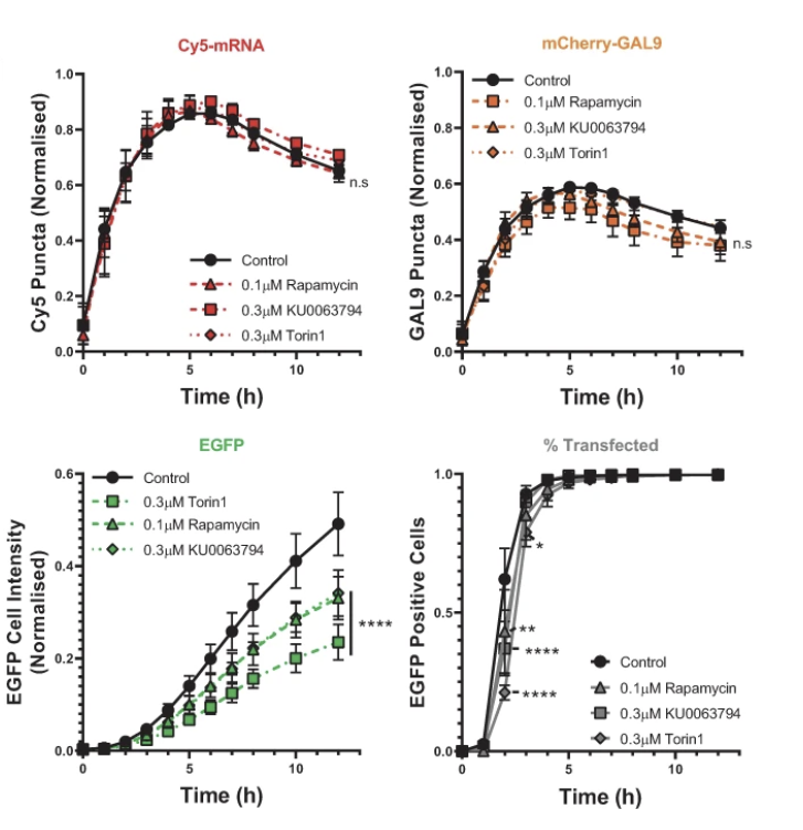
Figure 1. Effects of mTOR-pathway inhibition on labelled mRNA uptake (Cy5-mRNA), Galectin-9 recruitment, EGFP expression and the proportion of EGFP-positive cells. Uptake and the Galectin-9 signal were broadly similar across conditions, whereas EGFP expression was reduced, illustrating that uptake, escape-associated membrane disruption and functional protein output report different parts of the delivery process. Reproduced from Munson MJ et al., Communications Biology 2021, Fig. 3h. PMID: 33594247. Licensed under CC BY 4.0.
These assays also require careful interpretation. Galectin recruitment reports membrane damage associated with escape; it does not directly count intact mRNA molecules available to ribosomes. A protein reporter integrates delivery, translation and reporter turnover, while an irreversible recombination reporter records that functional delivery occurred rather than its continuing duration. PMID: 33594247; PMID: 30275336
For development, I would use these methods to test specific alternatives. If two LNPs produce different protein levels despite similar cellular uptake, the next experiment should help distinguish altered intracellular processing from altered RNA or protein kinetics. Repeating the same total-uptake measurement with more precision would leave that question unresolved.
The payload determines the useful bridge to pharmacology
A secreted protein can provide a relatively accessible connection between mRNA administration and activity. Once the protein enters circulation, its distribution, clearance and target interaction can often be represented using familiar biologics models. Its input is now synthesis and secretion by transfected cells, so the observed concentration-time profile combines protein production with protein disposition.
A 2026 study of BioNTech’s BNT141 and BNT142 illustrates this approach. These products encoded a full IgG and a bispecific T-cell engager, respectively. A translational model combined mRNA elimination and translation parameters with protein kinetics. Using non-human-primate data, the resulting predictions of human translated-antibody exposure were within twofold of the observed values. The example is useful because it shows how familiar protein PK can be connected to the less familiar kinetics of an mRNA input. PMID: 42035344
Recombinant-protein data can provide another useful constraint. In an academic modelling framework published by Campanile and colleagues, data from administered antibodies were used to estimate protein-specific parameters before fitting the additional mRNA-related processes. This separation helps address an otherwise difficult question: how much of a prolonged protein profile reflects continued synthesis, and how much reflects slow elimination? PMID: 42568897
Intracellular proteins require a different bridge. In the mRNA-3927 propionic-acidaemia programme, investigators linked plasma mRNA to hepatic propionyl-CoA carboxylase (PCC) protein through an effect compartment, then connected protein expression to circulating metabolic biomarkers. The effect compartment represented a delay between measured mRNA and protein response; it should not be interpreted as a directly sampled cytoplasmic compartment. PMID: 36577040
Transcription factors introduce further complexity. In our preclinical HNF4A work, functional assessment included gene-expression profiling, single-cell RNA sequencing and chromatin immunoprecipitation, with paraoxonase-1 (PON1) identified as a direct target contributing to the antifibrotic response. The work provides an experimental connection between expression and downstream function. For a quantitative model of such a therapy, nuclear activity and the resulting cellular response would need to be connected to the amount and distribution of expressed protein. PMID: 34453962
For such a programme, a useful model may need to distinguish how many relevant cells are affected from how strongly each cell responds. As a hypothetical example, the same mean tissue expression could arise from high expression in a small fraction of cells or moderate expression across many cells. If activity has a threshold or saturates within individual cells, those distributions can produce different tissue-level effects.
Use only as much mechanism as the decision requires
Existing pharmacological tools can represent much of this biology. Compartmental models describe measured disposition, effect compartments can accommodate delays, and turnover models connect protein production and loss to downstream responses. More elaborate models become useful when the question requires an explicit account of tissue distribution, intracellular processing or biological feedback.
The Campanile framework is an example of physiologically based pharmacokinetic modelling, or PBPK. It combined an established antibody-distribution model with a deliberately compact LNP-mRNA module and was calibrated and evaluated against five preclinical datasets. Several upstream processes were represented through lumped parameters rather than treating every intracellular rate as independently measurable. Useful as it is, this remains a preclinical framework rather than a human-validated model. PMID: 42568897
For vaccines, the relevant response extends through antigen presentation and immune-cell dynamics. A model developed by Dasti and colleagues linked molecular events and antigen-presenting cells to antibody responses, using published BNT162b2 antibody data for calibration and evaluation across regimens. This is quantitative systems pharmacology, or QSP: the model extends beyond the disposition of the administered material to represent interacting biological processes. Agreement with antibody measurements supports that output, without independently confirming every upstream mechanism or demonstrating protection against infection. PMID: 40420402
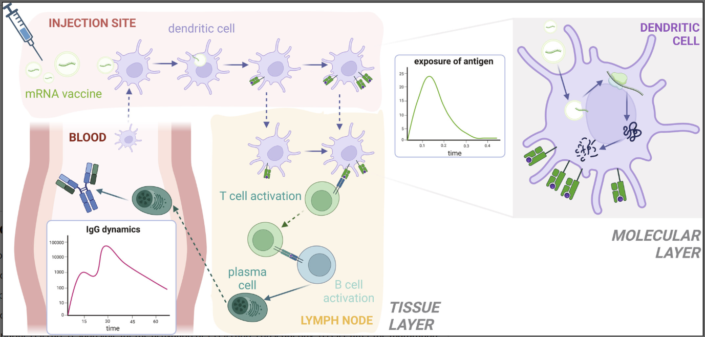
Figure 2. Multiscale QSP model structure for an mRNA vaccine, linking intracellular mRNA processing and antigen presentation with events at the injection site, draining lymph node and blood, and ultimately with antibody dynamics. The figure illustrates how the relevant model can extend well beyond disposition of the administered mRNA when the development question concerns an immune response. Reproduced from Dasti F et al., CPT: Pharmacometrics & Systems Pharmacology 2025, Fig. 1. PMID: 40420402. Licensed under CC BY 4.0.
The choice depends on the proposed use. A model intended to compare two delivery technologies needs evidence about the processes that distinguish those technologies. A model intended to describe antibody waning within an established regimen may be useful with a much more compressed representation of the upstream steps. A model’s detail should follow the prediction it needs to support.
A good fit does not prove the mechanism
Greater mechanistic detail introduces more quantities that need to be measured, estimated or assumed. When only a few outputs are observed, different parameter combinations can explain the same data. Identifiability analysis asks whether the available observations can distinguish those combinations, and separates limitations inherent in model structure from those caused by sparse or noisy measurements. PMID: 19505944
Consider a deliberately simple model in which protein production equals the amount of cytoplasmic mRNA multiplied by a translation-rate parameter. Doubling the assumed mRNA amount and halving the translation rate leaves protein production unchanged. If only the protein curve is measured, that curve cannot distinguish these two explanations without additional information.
This becomes consequential when the team is deciding what to optimise. A protein curve alone cannot establish whether limited expression reflects poor cytoplasmic availability or inefficient translation. Selecting an intervention directed at one of those processes requires additional evidence, even when the model describes the observed protein profile accurately.
Sensitivity analysis and identifiability analysis answer different questions. The first asks which parameters influence a chosen output; the second asks whether the observations can determine their values.
When a prediction will drive a development decision, I would also want to know whether alternative plausible parameter sets or model structures change the conclusion. Testing against data that were not used for fitting is particularly valuable when the intended use involves a new dose, schedule, formulation or population.
Align the assay with the model
This creates a close relationship between bioanalysis and modelling. If a model compartment represents intact, translation-competent cytoplasmic mRNA, total tissue RNA measured by a sequence-specific assay cannot automatically be assigned to that compartment. The measurement and the biological quantity need an explicit connection.
RT-qPCR detects a selected sequence region; detection alone does not establish that the entire transcript remains intact. General RNA-integrity studies have used comparisons between distant transcript regions to examine degradation. Those methods illustrate the distinction between detecting a sequence and assessing integrity, although they are not themselves validated assays of therapeutic mRNA translation competence. PMID: 29922590
Even the measured concentration can depend on the analytical workflow. A comparison of 77 clinical serum samples found that RT-qPCR yielded lower mRNA concentrations than branched-DNA analysis, with differences depending on sample preparation. PK parameters were nevertheless comparable in the small subset analysed. The practical lesson is that the assay method and its comparability need to be understood before concentration data are treated as interchangeable. PMID: 40903636
I would therefore define each important model observation in biological and analytical terms before finalising the study. What molecular species does the assay detect, in which matrix, and what interpretation depends on it? For protein assays, the corresponding question is whether the measurement reports total protein, a functional form or an activity, and whether endogenous protein complicates attribution to the administered mRNA.
Sampling can then be designed around the uncertainty that needs resolving. Early measurements may separate delivery and the onset of expression, while later measurements may help separate continued production from protein persistence and downstream response. Where repeated tissue sampling is impractical, the limitations of accessible surrogates should remain visible in the model.
An exposure metric also needs a biological rationale. Before reducing a profile to an area under the curve or a peak concentration, I would ask whether the intended response depends on that quantity, on time above a threshold, or on the fraction of cells expressing functional protein. Those possibilities lead to different sampling and modelling requirements.
The same discipline applies when the product changes. A plasma-mRNA relationship established for one formulation should be tested before being used to infer productive delivery after an LNP change. Agreement in an upstream measurement alone cannot establish agreement in the unobserved downstream process.
Start with the development decision
Before commissioning an mRNA PK/PD study, I would want the team to specify the decision the data are expected to support. Comparing delivery systems, understanding a lack of response, selecting a dosing interval and evaluating a different patient population can require different measurements and different levels of model detail.
For each decision, the programme should identify the relevant exposure, the PD readout that tests its biological consequence, and the assumptions connecting the two. The resulting model should make clear which quantities were measured directly, which were inferred, and which remain insufficiently constrained.
That is the practical value of distinguishing distribution, productive intracellular exposure and effector exposure. It allows the programme to collect evidence that connects the administered product to its intended biological action, while keeping the limits of that evidence explicit.